Navigational bronchoscopy is an image guided bronchoscopic procedure that uses electromagnetic technology and allows physicians to reach deep into the lung and biopsy small nodules previously inaccessible to conventional bronchoscopy.
Prior to performing the procedure, the computer creates a virtual map using patients latest CT chest. This map is then used to plan the best route to reach the lesion. Navigation to the target lesion is performed using steerable flexible instruments and GPS-like technology.
“This has significantly improved our ability to reach small nodules in the periphery of the lung” says Dr Shostak, Interventional Pulmonologist and Assistant Professor of Medicine in Cardiothoracic Surgery.
Despite these improvements, electromagnetic navigational bronchoscopy still has some drawbacks, including “CT-to-body divergence” – a discrepancy between the location of a nodule as seen on pre procedure scan and the actual location of the nodule during the procedure. This has resulted in a decrease in diagnostic yield, especially for nodules that are small.
Going beyond traditional navigational bronchoscopy
Here at Weill Cornell Medicine/NewYork-Presbyterian Dr. Shostak is one of the few bronchoscopists in New York City that uses the Fluroscopic Navigational Platform. This upgraded system goes beyond traditional navigational technology by providing improved GPS-style guidance and technological upgrades to overcome challenges associated with CT-to-body divergence.
“The system significantly improves our ability to reach smaller nodules and diagnose lung cancer early” says Dr Shostak.

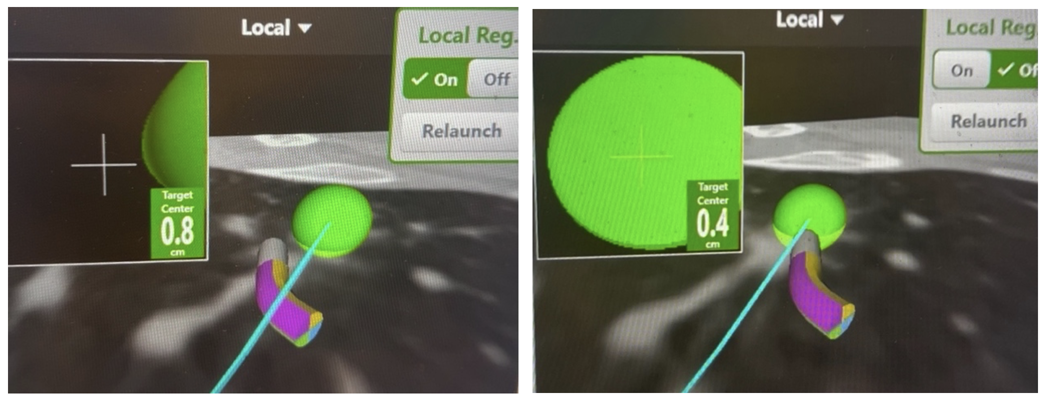
(Right). Catheter pointing at the virtual target represented by the green ball, suggesting that the instrument is in the center of the lesion. (Left). the Fluoroscopic Navigational System corrects for CT-to-body divergence demonstrating that the catheter is not in the lesion as previously suggested. Ability to perform this correction greatly improves the diagnostic accuracy of the procedure
By correcting for the CT-to-body divergence, the Fluoroscopic Navigational System significantly increases ability to reach and biopsy small peripheral nodules with better accuracy, allowing pulmonologists to diagnose lung cancer earlier.


