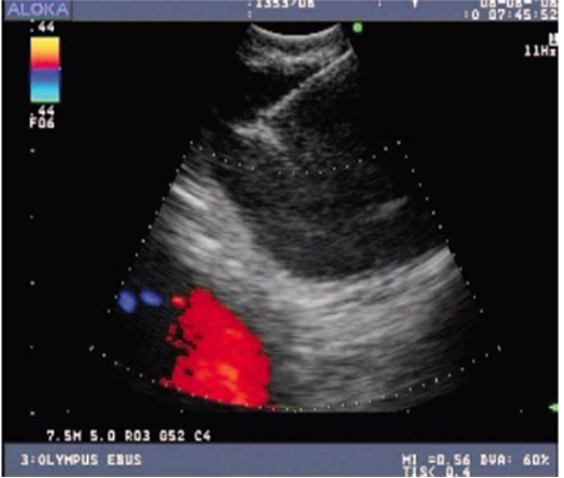
Bronchoscopy with Endobronchial Ultrasound demonstrating insertion of a needle into a lymph node performed under ultrasound guidance. Doppler allows for visualization of nearby blood vessels.
Enlargement of lymph nodes in the chest can result from a variety of conditions, ranging from benign disorders such as sarcoidosis, to more serious conditions such as lymphoma and lung cancer.
“At Weill Cornell Medicine | NewYork-Presbyterian Hospital we routinely perform endobronchial ultrasound guided biopsies which enables us to accurately diagnose various disorders affecting lymph nodes in the chest using a small camera without need for surgery.” says Dr. Eugene Shostak, Interventional Pulmonologist and Assistant Professor of Medicine in Cardiothoracic Surgery.
Endobronchial ultrasound consists of a tiny ultrasound probe attached to the tip of the flexible bronchoscope, which permits visualization of lymph nodes located outside the airway passage and are therefore not visible during regular bronchoscopy.
Endobronchial ultrasound procedure is often combined with either navigational bronchoscopy or robotic bronchoscopy procedures to collect most information during same procedure. In cancer patients this can in turn decrease the time from diagnosis to initiation of treatment.
Dr. Shostak adds, “we try to avoid any delays in care by offering a one-stop shop model, where we combine endobronchial ultrasound with navigational or robotic assisted bronchoscopic procedure and perform sampling of both lymph nodes and lung nodules in the same setting”
Weill Cornell Medicine Cardiothoracic Surgery
525 East 68th Street
Box 110
Suite M 404
New York, NY 10065
Directions
Phone: (212) 746-5166
Email: ctsurgery@med.cornell.edu